3. Visceral adipose tissue (VAT) analysis
·Visceral adipose tissue is adipose surrounding internal organs (TAG 5- Yellow -150 to -50 HU)
VAT can be analyzed using either the paint function or the Grow 2D function (Grow 2D works best with large amounts of VAT)
|
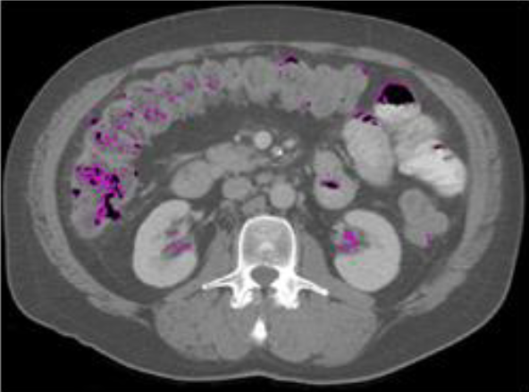
What is not considered VAT: ·Fat inside the kidney or liver ·Fat inside the intestines (this is fat from food, not VAT) (this is inside the black spaces which are air pockets in the intestine)
|
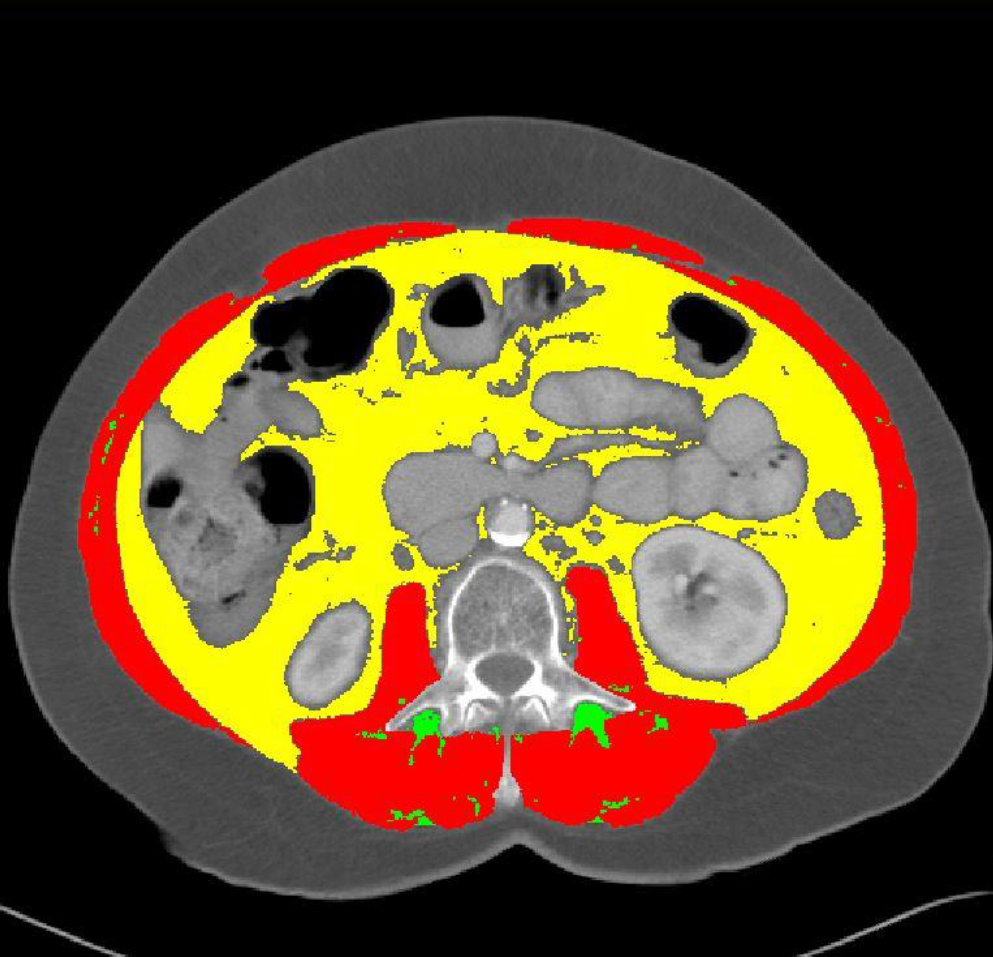
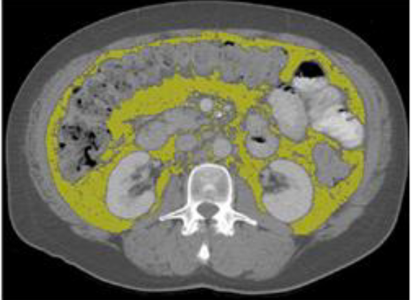
What is considered VAT: ·Area between the psoas and vertebra ·Note about analysis of VAT surrounding the intestines: color all the way to the intestines so that it is touching the grey border, but do not color inside the grey border
|
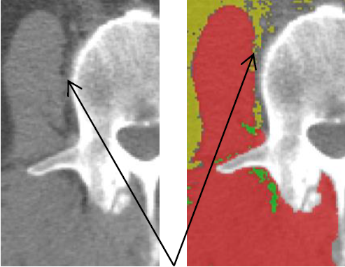
Make sure to analyze area between psoas muscle and vertebra as VAT
VAT analysis checklist
·Fat inside the kidneys is not analyzed
There should be no VAT in the kidneys as this is perirenal fat and not visceral fat.
·Fat inside the intestines are not analyzed
There should be no VAT inside the intestines because this is fat from food and not VAT (i.e., there should be no fat inside the black holes)
·Fat inside the liver is not analyzed
·Are the VAT areas near/bordering the muscle colored?
The fat found immediately between the vertebra and the psoas muscles should be analyzed as VAT
·Are the VAT areas bordering the intestines colored?
This is all the way up to the grey line surrounding the black holes
·Are the VAT areas near the vertebra analyzed?
·Are the VAT areas between the kidney and psoas muscle analyzed?