Landmarking is the process of identifying the anatomical location of interest for analysis. The 3rd lumbar vertebra (L3) is the landmark of interest.

Anatomical knowledge of the vertebral column will be used to landmark.
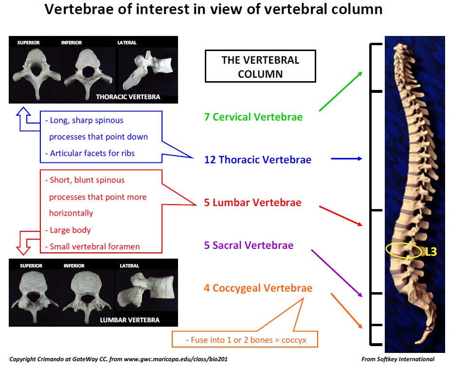
Three key anatomical features to remember when searching for the L3 landmark on CT images
• 12 thoracic vertebrae and 5 lumbar vertebrae
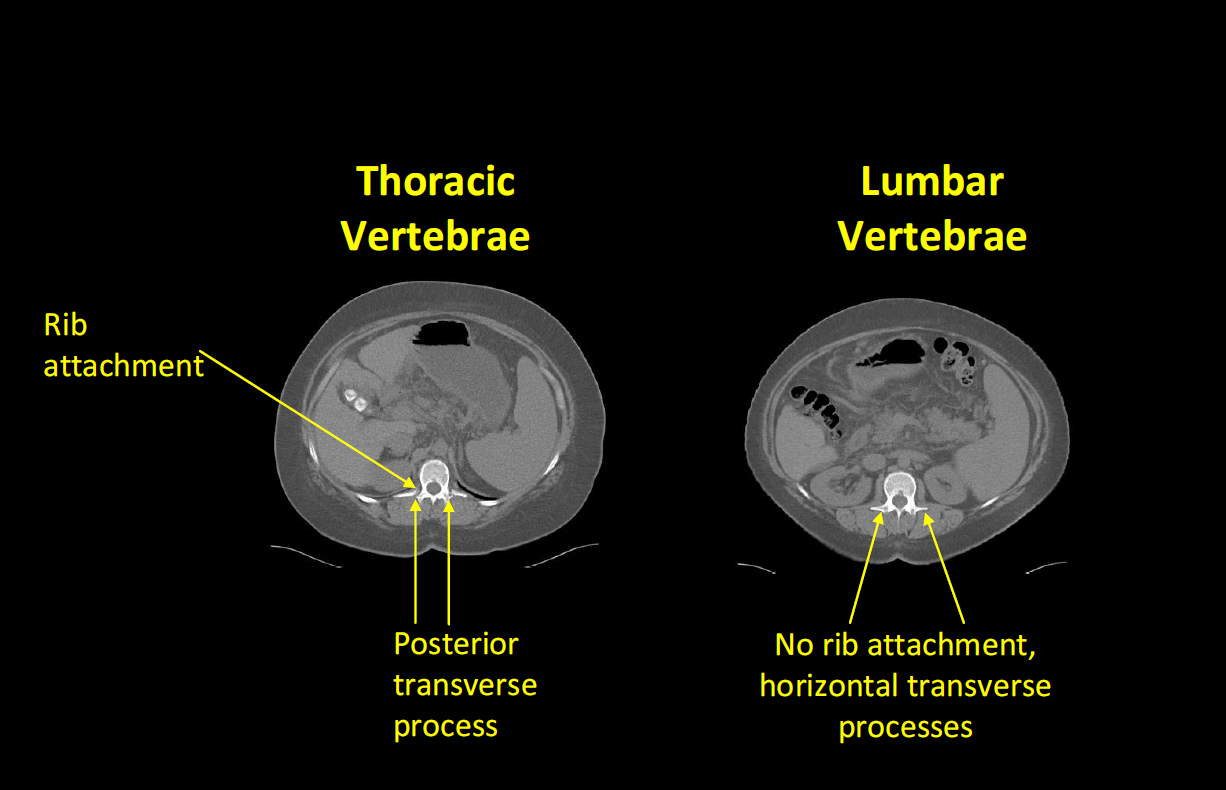
• Thoracic vertebrae have rib attachments, lumbar vertebrae do not
• Thoracic vertebrae have transverse processes that point more posterior (towards the back); lumbar vertebrae have transverse processes that point more towards the sides (or horizontally)
Landmarking Steps
1. Identify the 1st lumbar vertebrae (L1)
·This will be the first vertebrae without a rib attachment
·It will also have transverse processes that point more horizontally (and therefore towards the left and right sides) than posterior (towards the back) (like the 12th thoracic vertebra)
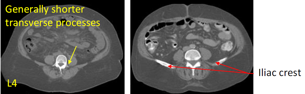
2. Once L1 has been found, use the transverse processes (TP) to count inferiorly (or downward) to L3. As you scroll down from L1, you will see that the TP disappear and reappear. Each time they disappear and reappear, you are at a different and lower vertebra.
·If you see the iliac crest (top of the pelvis) or short transverse processes and a wider vertebral body, you may be at L4.
3. Once L3 has been found, choose the image with both transverse processes clearly visible. This will be right in the middle of L3.

Important note: as seen in the previous figure, each vertebra encompasses several CT images, not only one image.
Landmarking tips
Other important bone structures when landmarking L3 CT images:
·Iliac Crest: The iliac crest is usually not visible at L3. If it is visible at the possible L3 image, count again to ensure that it is not at L4.
·Ribs: There are usually no ribs showing close to the obliques and transversus abdominus muscles at L3. If there are ribs on the possible L3 image, check again to ensure that the image is not from L2 or L1.

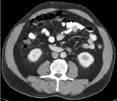
T12, L1, L2, L3, and L4 CT images